Versiti - Image Gallery | Tirthadipa Pradhan-Sundd Laboratory | Versiti Blood Research Institute
Explore our service lines

Image Gallery

Research images from the Pradhan-Sundd laboratory at Versiti Blood Research Institute.

Liver Intravital Imaging

Intravital imaging of WT mouse liver showing movement of blood (FITC -dextran) through liver sinusoids.

Intravital imaging of WT mouse liver showing movement of blood (red; TXR-Dextran) ) and bile (green, CF) in real time.

Intravital imaging of mouse liver showing movement of blood (red) and bile (green) in real time.

WT mouse liver showing movement of blood through sinusoidal vessels (Texas-Red) and Neutrophils (blue).

SCD mouse liver showing vasoocclusion at baseline.

Ferritin (green) and F4/80 (red) in mouse liver section

Ferritin (green) and F4/80 (red) in mouse liver section.

Intravital imaging of sickle cell mouse liver . In red Texas red dextran and Carboxyflurescein is in green. Circled area shows vasoocclusion.

Intravital imaging of sickle cell mouse liver . In red Texas red dextran and Carboxyflurescein is in green. Circled area shows vasoocclusion.

Intravital imaging of sickle cell mouse liver. In red Texas red dextran and Carboxyflurescein is in green. Circled area shows vasoocclusion.

Intravital imaging of sickle cell mouse liver. In red Texas red dextran and Carboxyflurescein is in green. Circled area shows vasoocclusion.

E-cadherin staining (green) in liver

E-cadherin staining (green) in liver.

in-situ hybridization@liver

in-situ hybridization@liver

in-situ hybridization@liver

in-situ hybridization@liver

Phalloidin staining in liver tissue

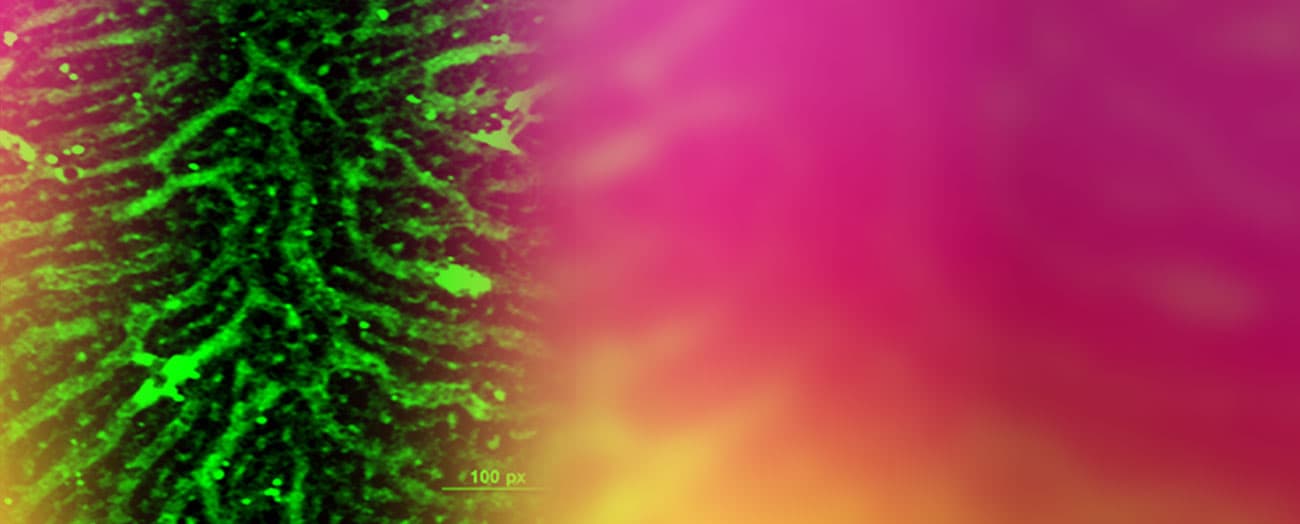
Phalloidin staining in liver tissue.

Volumetric analysis of sinusoidal structure

Volumetric analysis of sinusoidal structure.

TIRF imaging of claudin in liver tissue

TIRF imaging of claudin in liver tissue.

Liver sinusoidal blood vessels as seen using multiphoton microscopy.

Liver sinusoidal blood vessels as seen using multiphoton microscopy.

E-cadherin in Hep3B cells.

E-cadherin in Hep3B cells.

Pradhan-Sundd Lab Image