
Versiti Blood Research Institute Articles
Blood Research Institute Investigator Wins Journal Cover Photo Contest

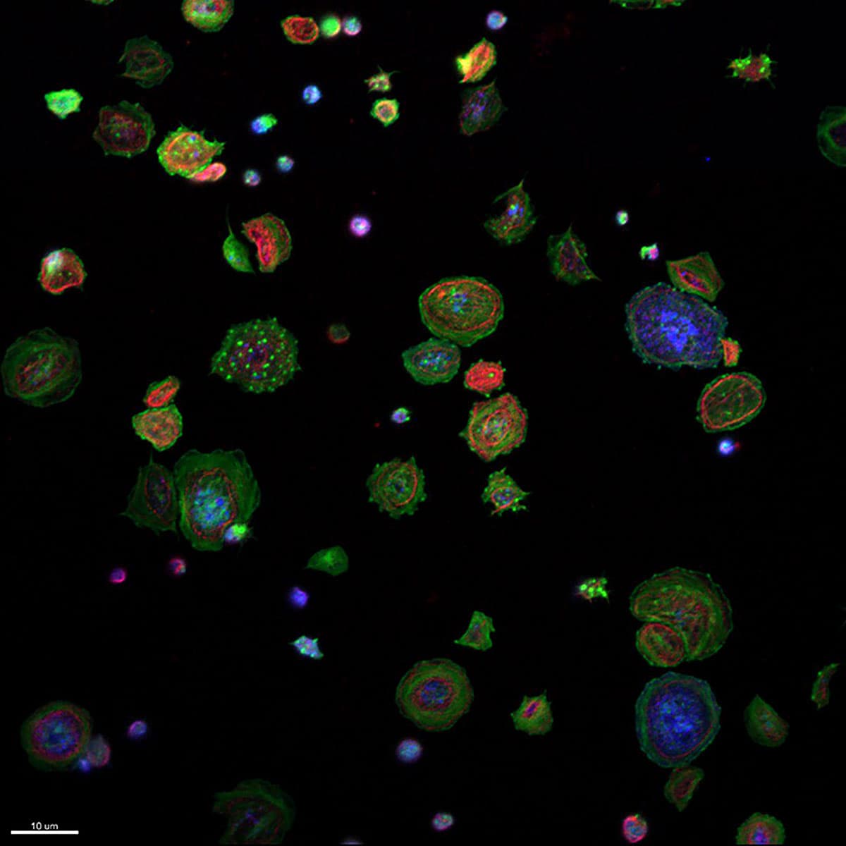
This particular image depicts platelets spreading on an artificial fibrinogen-coated surface, demonstrating what would happen if someone cut themselves. The amount of blood is tiny; one drop of blood typically yields millions of platelets, but this image shows about 50. To the untrained eye, it looks like fireworks in a night sky, but to an expert like Dr. Falet, something appears abnormal.
Each color is a different protein that can be found in or on the surface of platelets:
- Green: filamentous actin, the most abundant protein in platelets and the major constituent of the platelet cytoskeleton.
- Red: tubulin, another constituent of the platelet cytoskeleton, polymers of which form a marginal ring in resting platelets, contributing to their distinctive discoid shape.
- Blue: the von Willebrand factor receptor, GPIbα, which plays a critical role in platelet adhesion at arterial shear rates.
The Versiti Blood Research Institute provides a wealth of imaging resources to its investigators. But for now, Dr. Falet is pleased that his image - which he views as a work of art - is being recognized. “It’s interesting to me that, in something so small as platelets, you can see the patterns that you see in the sky - the art of it,” he says.
About the expert: Herve Falet, PhD, is an investigator at the Versiti Blood Research Institute and assistant professor in the Department of Cell Biology, Neurobiology and Anatomy at the Medical College of Wisconsin.